facs flow cytometry protocol
Discriminate between live and dead cells during flow cytometry. 14 bright fixable dyes.

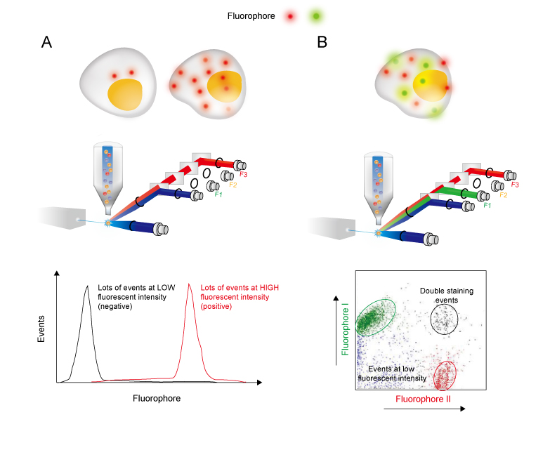
Schematic Representation Of The Flow Cytometry Protocol Download Scientific Diagram
Ad Bright and highly specific fixable dead cell stain for flow cytometry.

. Recombinant proteins designed for biological medicine RD. GENERAL CELL STAINING PROTOCOL FOR FLOW CYTOMETRY 1 Except for cells grown in culture cells obtained directly from tissues must first be resolved to a single cell suspension. Protocols are available for.
This incubation must be done in the dark. The flow cytometry protocols below provide detailed procedures for the treatment and staining of cells prior to using a flow cytometer. Can Remove Approximately 50 Million Cells.
Talk To A Scientist Today. Flow Cytometry FACS Protocols PSR The BD FACSCalibur platform allows users to perform both cell analysis and cell sorting in a single benchtop system. Use Microbubbles For Gentle Cell Separation.
Can Remove Approximately 50 Million Cells. Super Bright Staining Buffer protocol. Explore protocols for sample preparation of mouse and rat leucocytes indirect staining of mononuclear cells reducing nonspecific staining with Fc Block intracellular cytokine staining.
Harvest wash the cells and adjust cell suspension to a concentration of 1-5 x 10 6 cellsmL in. The following flow cytometry. Ad High Specificity For Cell Separation.
Ideal Shipping Method According To Items Temperature Requirement. Cell Preparation for Flow Cytometry Protocols Invitrogen eBioscience reagents Red Blood Cell Lysis Protocols. As cells scatter laser light in.
Flow cytometry is the measurement of chemical and physical properties of cells as they flow one by one through an integration point most commonly a laser. Primary Antibody Staining 1. Flow cytometry FACS staining protocol Cell surface staining Harvest wash the cells single cell suspension and adjust cell number to a concentration of 1-5x10⁶ cellsml in ice cold FACS.
The system supports a wide. Indirect labelling requires two incubation steps. With Fluidigm Maxpar Pathsetter Software Data Analysis Can Be Done in Minutes.
Ad Transformative - run difficult samples with a system that is less sensitive to clogging. Rethink flow cytometry with more sensitivity and more accurate detection. The following flow cytometry staining protocol.
By staining cell surface markers researchers can identify specific cell populations and perform fluorescence-activated cell sorting FACS. Ad Bring the Power of Cytometry to Advancing Infectious Disease Research. Use Microbubbles For Gentle Cell Separation.
If you are unable to immediately read your samples on a cytometer keep them shielded from light and in. Indirect flow cytometry FACS protocol General procedure for flow cytometry using a primary antibody and conjugated secondary antibody. General procedure for flow cytometry using a conjugated primary antibody.
Add 1 μg of primary antibody. Request a quote and see how Agilent has advanced the boundaries of flow cytometry. General protocols for flow cytometry.
Flow cytometry was performed on a BD FACScan flowcytometry system. Wash the cells 3 times by centrifugation at 400 g for 5 min and resuspend them in ice. Cell Surface Staining of Human PBMCs and Cell Lines.
Perform fluorescence activated cell sorting FACS or flow cytometric analysis. Ad Fpf flow cytometry derived from HEK293 high Purity high batch-to-batch consistency. Rethink flow cytometry with more sensitivity and more accurate detection.
Ad Used To Preserve Cell Surface Epitopes That Have Previously Been Stained. Direct staining of cells. Incubate for at least 20-30 min at room temperature of 4C.
Flow Cytometry is used for research applications such as immunophenotyping DNA studies cell cycle analysis and fluorescence-activated cell sorting FACS. Ad High Specificity For Cell Separation. Ad Agilent NovoCyte flow cytometers are built to provide high data quality and flexibility.
Ad Transformative - run difficult samples with a system that is less sensitive to clogging. Talk To A Scientist Today.
Flow Cytometry Guide Creative Diagnostics

Protocol For Renal Cells Isolation And Macrophage Detection By Flow Download Scientific Diagram

Flow Cytometry Facs Protocols Sino Biological

Workflow For Establishing The Fit For Purpose Of A Flow Cytometry Download Scientific Diagram

Fluorescence Activated Cell Sorting Facs Sino Biological

Optimized Flow Cytometric Protocol For The Detection Of Functional Subsets Of Low Frequency Antigen Specific Cd4 And Cd8 T Cells Sciencedirect
Intranuclear Immunostaining Based Facs Protocol From Embryonic Cortical Tissue Sciencedirect

Flow Cytometry Creative Biolabs

Flow Cytometry Protocols